
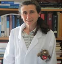
Equipe Equipe Presage
caroline.fonta@cnrs.fr
Tél : 06 62 74 61 44

My primary research project focuses on identifying markers—or combinations of markers—that characterize and predict brain aging using a non-human primate model (marmoset monkey). These markers are primarily derived from non-invasive brain imaging techniques, combined with cognitive performance assessments, in collaboration with Pascal Girard at CerCo.

⦁ Kennel P, Fonta C, Guibert R, Plouraboué F. 2017. Analysis of vascular homogeneity and anisotropy on high-resolution primate brain imaging. Human Brain Mapping 38(11):5756-5777. doi: 10.1002/hbm.23766
⦁ Risser L, Sadoun A, Mescam M, Strelnikov K, Lebreton S, Boucher S, Girard P, Vayssière N, Rosa MGP, Fonta C. (2019). In vivo localization of cortical areas using a 3D computarized atlas of the marmoset. Brain Structure and Function 224(5):1957-1969. doi: 10.1007/s00429-019-01869-x
⦁ Rodriguez-Callejas JDD, Cuervo-Zanatta D, Rosas-Arenallo A, Fonta C, Fuchs E, Perez Cruz C. 2019. Loss of ferritin-positive microglia relates to increased iron, RNA oxidation and dystrophic microglia in brain of aged male marmoset. American Journal of Primatology e22956.doi: 10.1002/ajp.22956.
⦁ Sadoun A, Rosito M, Fonta C, Girard P. 2019. Key periods of cognitive decline in a non-human primate model of cognitive aging, the common marmoset (Callithrix jacchus). Neurobiology of Aging 74:1-14. doi: 10.1016/j.neurobiolaging.2018.10.003
⦁ Arribarat G, Mescam M, Desmoulin F*, Fonta C*. 2025. Arterial and venous flow dynamics are modified by age in the non-human primate. Imaging Neuroscience, Volume 3, 2025. https://doi.org/10.1162/IMAG.a.66.
I also address the functions, in the brain, of TNAP, a tissue- and substrate-nonspecific ectoplasmic phosphatase, historically associated with bone mineralisation and liver pathophysiology. We mapped in time and space TNAP activity and expression in mouse (WT and TNAP-KO) and primate brains. We developed a transdisciplinary approach to analyse metabolomic consequences of TNAP inhibition or acute/chronic systemic inflammation. I also collaborate with Lionel Nowak (CerCo) to assess the ectonucleotidase function of TNAP in synaptic transmission. (6,7,8)


⦁ Cruz T, Gleizes M, Balayssac S, Mornet E, Marsal G, Millán JL, Malet-Martino M, Nowak LG, Gilard V, Fonta C. 2017. Identification of altered brain metabolites associated with TNAP activity in a mouse model of hypophosphatasia using untargeted NMR-based metabolomics analysis. J Neurochem 140:919-940. doi-org-s.docadis.univ-tlse3.fr/10.1111/jnc.13950
⦁ Perrier S, Gleizes M, Fonta C, Nowak LG. Effect of adenosine on short-term synaptic plasticity in mouse piriform cortex in vitro: adenosine acts as a high-pass filter. Physiological Reports, 2019, 7 (3), pp.e13992. ⟨10.14814/phy2.13992⟩.
⦁ Goettsch C, Strzelecka-Kiliszek A, Bessueille L, Quillard T, Mechtouff L, Pikula S, Canet-Soulas E, Millan JL, Fonta C, Magne D. (2020). TNAP as a therapeutic target for cardiovascular calcification – a discussion of its pleiotropic functions in the body. Cardiovasc Res. Oct 18;cvaa299. doi: 10.1093/cvr/cvaa299.
⦁ Bessueille L, Briolay A, Como J, Mebarek S, Mansouri C, Gleizes M, El Jamal A, Buchet R, Dumontet C, Matera EL, Mornet E, Millan JL, Fonta C, Magne D. (2020). Tissue-nonspecific alkaline phosphatase is an anti-inflammatory nucleotidase. Bone. 133:115262. doi: 10.1016/j.bone.2020.115262
⦁ Gleizes M, Fonta C*, Nowak L*. (2022) Inhibition of ectonucleotidases has paradoxical effects on synaptic transmission in the mouse cortex. J Neurochem 160:305-324. doi: 10.1111/jnc.15558.
⦁ Book : Caroline Fonta and László Négyessy (Eds) 2015. Neuronal Tissue-Nonspecific Alkaline Phosphatase (TNAP), Subcellular Biochemistry, Vol. 76. Springer ISBN 978-94-017-7196-2, doi: 10.1007/978-94-017-7197-9. 350pp; book.
I contribute to a paleoneurobiology project led by Amélie Baudet (10). Grooves visible on endocranial surfaces serve as important markers of functional brain areas in humans. Atlases of these sulci, along with their probability maps, help document variations in sulcal patterns across the cerebral cortex. By comparing such atlases from both human and non-human primate endocrania, we can analyze hominin fossil skulls to better understand the evolution of the cerebral cortex. https://www.endomap.org/ (9,10)

⦁ De Jager EJ, Van Schoor AN, Hoffman JW, Oettle AC, Fonta C, Mescam M, Risser L, Beaudet A (2019). Sulcal pattern variation in extant human endocasts. Journal of Anatomy 235(4):803-810. doi.org/10.1111/joa.13030.
⦁ Beaudet A, Fonta C. (2020) Aux origines de l’humanité : retracer les grandes étapes de notre évolution biologique et culturelle à partir de l’étude du cerveau de nos ancêtres. Intellectica, 2020/2, Emergence et évolution de la Cognition, ed S.A de Beaune, 73:93-106.https://hal.archives-ouvertes.fr/hal-03165140v1
⦁ de Jager EJ, Risser L, Mescam M, Fonta C, Beaudet A. (2022). Sulci 3D mapping from human cranial endocasts: A powerful tool to study hominin brain evolution. Hum Brain Mapp 43:4433–4443. doi: 10.1002/hbm.25964
⦁ Caroline Fonta, Amélie Beaudet, Annael Le Poullennec. BRAIN EVOLUTION: PALAEOSCIENCES, NEUROSCIENCE AND ARTIFICIAL INTELLIGENCE. Lesedi : Carnets de terrain = Lesedi : Field notes, 26, 44 p., 2024, LESEDI. ⟨hal-04785753⟩
Main external collaborators
- Franck Desmoulin (Tonic INSERM 1214, Toulouse)
- Jean Pierre Raymond, Michel Fournié (Institute of Mathematics, Toulouse)
- Moktar Zagzoule, Patricia Cathalifaud (Institute of Fluid Mechanics, Toulouse)
- Stéphanie Salmon, Nicolas Passat (University of Reims)
- Olivier Baledent (Chimere, University of Amiens)
- Véronique Gilard, Fabrice Collin, Stéphane Balayssac (IMRCP, University of Toulouse)
- David Magne, Anne Briolay (ICBMS, University of Lyon)
- Jose Luis Millan (Burham Institute, La Jolla, CA, USA
- Laurent Risser (Institute of Mathematics, Toulouse)
- Amélie Beaudet (PALEVOPRIM, University of Poitiers)